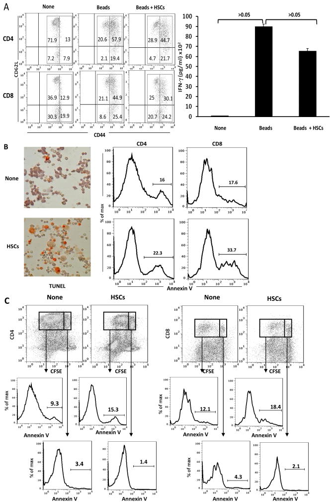
Figure 3. HSCs inhibit T cell response via induction of B7-H1-mediated apoptotic death.
Irradiated human HSCs were added (at a ratio of 1:10) at the beginning into a one-way MLR culture in which the proliferation of PBMC-derived T cells was elicited by anti-CD3/CD28 beads. (A) HSCs do not suppress T cell activation. Expression of CD44 and CD62L were analyzed on naïve CD4 and CD8 T cells (None), stimulated by anti-CD3/CD28 beads or plus addition of HSCs by flow cytometry. The concentration of IFN-γ in supernatant was measured by CBA assay. Naïve T cells expressed intermediate levels of CD44. TCR stimulation markedly enhanced expression of CD44, but modestly inhibited expression of CD62L on both CD4 and CD8 T cells, and markedly increased IFN-γ production. Addition of HSCs did not affect these parameters of activation. The number is percentage of positive cells in each gated area. (B) HSCs induce T cell apoptosis. Apoptosis of the T cells were determined by staining of TUNEL (cytospin histochemistry), as well as by expression of annexin V on T cells (flow analysis gated in CD3+ cells). The number is percentage of annexin V+ T cells. (C) HSCs preferentially induce apoptosis in activated T cells. Expression of annexin V was analyzed in dividing and non-dividing CD4 and CD8 T cells. The number is percentage of annexin V+ cells.

