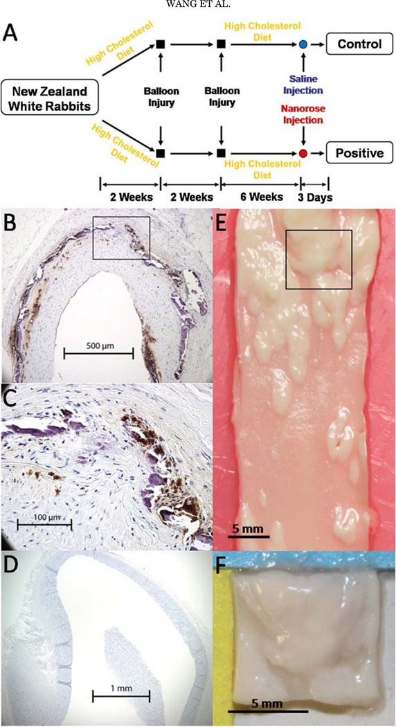
Fig. 2.
A: Rabbit model of aorta inflammation with intimal hyperplasia. B: RAM-11 staining of a cross-section of abdominal aorta. C: Amplified view of the black box in (B). Brown color indicates macrophages. D: RAM-11 staining of a cross-section of thoracic aorta. E: A piece of abdominal aorta with intimal hyperplasia. F: An aorta segment (8×8×2 mm) cut from the black box in (E).

