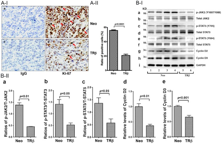
Figure 4.

Inhibition of cell proliferation by TRβ in tumors derived from MCF-7-TRβ cells. A-I: Immunohistochemical analysis of protein abundance of the nuclear proliferation marker, Ki-67, in tumors. Sections of tumors derived from MCF-7-Neo cells (panels a and b) and MCF-7-TRβ cells (panels c and d) were treated with control anti-IgG (panel a and c) or with anti-Ki-67 antibodies (panels b and d) as described in Materials and Methods. The Ki-67 positively stained cells are indicated by arrows. A-II: The Ki-67-positive cells were counted and expressed as percentage of Ki-67-positive cells versus total cells. The data are expressed as mean ± SE (n=3). B-I: Western blot analysis of key regulators and effectors in JAK2-STAT signaling pathway in tumors. Tumors were excised from the injection sites (hind flanks) of athymic nude mice, and the Western blot analysis was carried as described in Materials and Methods. Lanes 1-4 were key regulators and effectors detected in extracts from tumors derived from MCF-7-Neo cells, and lanes 5-6 were from tumors derived from MCF-7-TRβ cells. The proteins (p-JAK, total JAK, p-STAT3, total STAT3, p-STAT5, total STAT5, cyclin D2, cyclin D3, and GAPDH) detected are as marked. B-II: The band intensities of the proteins detected in B-I were quantified and compared. The data are shown as mean ± SE (n=2-4). The p values are shown.
