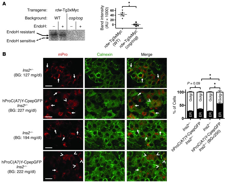
Figure 6. Rescue of mutant Tg and blockade of WT proinsulin in primary tissue from animal models of disease.
(A) Lobules of thyroid glands were freshly prepared from mice of the indicated genotypes. Secretory proteins delivered for posttranslational iodination were labeled by incubation of thyroid lobules with 1.0 μCi/μl Na125I for 30 minutes, as described in Methods. The thyroid lobules were then lysed and immunoprecipitated with anti-Myc. The immunoprecipitates were either mock-digested or digested with EndoH, as in Figure 2B, and then analyzed by SDS-PAGE and autoradiography. *P < 0.05. (B) Pancreata from 6-week-old mice, with the genotypes indicated, were fixed in paraffin, sectioned, deparaffinized, and immunostained with antibodies specific to mPro (red) and calnexin to mark the ER (green). From confocal microscope images (scale bar: 10 μm), a blinded reader scored the localization of WT mPro in each β cell as either a predominant juxtanuclear crescent of increased intensity (Golgi, consistent with previous reports, refs. 20, 23; e.g., see arrows) or mainly colocalized with calnexin (ER; e.g., see arrowheads). Quantitation of these data is shown as mean ± SEM from n = 5 mice with 5 islets per mouse. BG, blood glucose. *P < 0.05.

