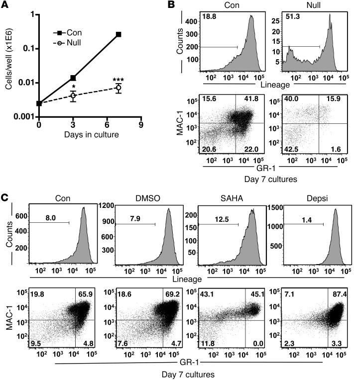
Figure 6. Inactivation of Hdac3 impairs progenitor cell proliferation differentiation.
(A) 2,500 WT or Hdac3-null LSK/FLT3– bone marrow cells were cultured on an OP9-GFP stromal layer in media containing IL-6, SCF, and LIF, and proliferation was monitored by counting the total number of cells 3–7 days after harvesting the bone marrow cells (day 3, P = 0.026; day 7, P ≤ 0.001; n = 3). (B) Flow cytometric analysis of day-7 cultures (from A) using a combination of anti-CD3, anti-B220, anti–GR-1, anti–MAC-1, and anti-Ter119 to distinguish mature cells from immature progenitor cells (top panel) or a combination of anti–GR-1 and anti–MAC-1 to quantify myeloid cell differentiation (bottom panel). (C) Flow cytometric analysis of WT LSK cells treated with DMSO, SAHA, or depsipeptide (Depsi) and cultured for 7 days as in (A). Representative plots show a combination of anti-CD3, anti-B220, anti–GR-1, anti–MAC-1, and anti-Ter119 to distinguish mature cells from immature progenitor cells (top panel) or a combination of anti–GR-1 and anti–MAC-1 to quantify myeloid cell differentiation (bottom panel). The numbers in each box indicate the relative percentage of the cells in the indicated gated population.

