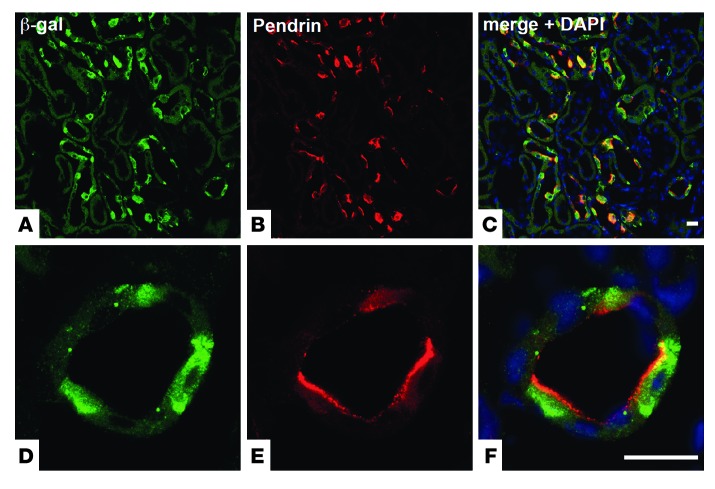
Figure 1. OXGR1 is expressed in type B and non-A–non-B intercalated cells of the CNT and the CCD.
(A) Immunostaining with anti–β-galactosidase antibody (green) shows that OXGR1 is expressed in a subset of cells in the renal cortex. (B) Immunolocalization of pendrin (red) in the apical membrane of type B and non-A–non-B intercalated cells. (C) Coimmunostaining with anti–β-galactosidase and anti-pendrin antibodies demonstrates that OXGR1 and pendrin are expressed in the same cells. Higher-magnification views of images in A–C are shown in D–F, respectively. Nuclei (DNA) are stained with DAPI (blue, C and F). Scale bars: 50 μm.

