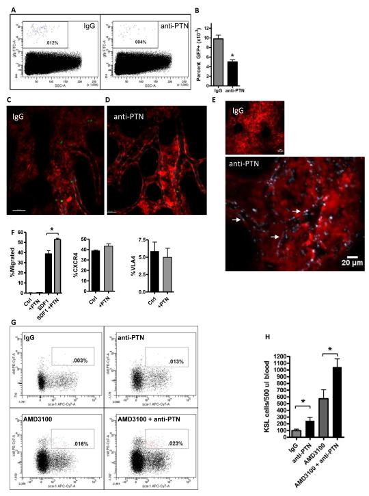
Figure 4. PTN Regulates the Homing and Retention of BM HSPCs.
(A) Systemic administration of anti-PTN significantly decreased HSPC homing to the BM niche. Representative FACS analysis is shown of percentage GFP+ donor hematopoietic cells in the BM of recipient C57Bl6 mice at 18 hours following intravenous infusion of BM Sca-1+lin−GFP+ cells after pre-treatment of recipient mice with either anti-PTN or IgG. (B) Anti-PTN treated mice contained > 2-fold decreased donor GFP+ cells in the BM at 18 hours post-infusion compared to IgG-treated control mice. n=5-6/group, means ± SEM, *p=0.0004. (C, D) Administration of anti-PTN inhibits the lodgment and transmigration of HPCs from the BM vasculature into the stem cell niche. Representative intravital images are shown of the calvarial BM space in living dsRed mice during the first 4 hours post-intravenous infusion of 3 × 10⁶ BM lin−GFP+ cells. Mice that were pre-treated with IgG antibody (C) demonstrated abundant homing of transplanted cells (green) from the BM sinusoidal vasculature (gray) into the niche, whereas mice pre-treated with anti-PTN displayed a markedly decreased number of donor HPCs within the extravascular BM space (D). A single GFP+ (green) cell is shown within the BM vasculature in the anti-PTN treated mice. See also Movies S1 and S2 for dynamic imaging of donor GFP+ hematopoietic progenitor cell homing in the BM of IgG-treated and anti-PTN treated mice. (E) Systemically administered anti-PTN binds extensively to the BM sinusoidal vasculature. Representative intravital imaging is shown of the calvarial BM of dsRed mice at 30 minutes after intravenous injection of either IgG-DyLight650 antibody (left, 20×, scale bar 50 μm) or anti-PTN-DyLight650 (middle, 20X, scale bar 50 μm). Binding and illumination of the BM sinusoidal vasculature by anti-PTN antibody (gray outline, white arrows) is shown in high power at right (4 times zoom of white box area in previous image, scale bar 20 μm). (F) Pre-incubation with PTN augments HPC migration to an SDF1 gradient. BM ckit+lin− cells demonstrated no migration to media alone or PTN in a 4 hour transwell assay (left, n=4/group). A percentage of BM ckit+lin− cells migrated to an SDF1 gradient in the lower chamber of transwell cultures. BM ckit+lin− cells that were pre-incubated with PTN × 1 hour demonstrated significantly increased migration to SDF1 compared to control cultures at 4 hours in transwell assay. n=6/group, means ± SEM, *p=0.002 (left). Incubation of BM ckit+lin− cells with PTN had no effect on cell surface expression of CXCR4 (middle) or VLA4 (right); n=6/group, means ± SEM. (G) Administration of anti-PTN promoted the rapid mobilization of HSPCs in wild type mice. Representative FACS plots are shown of the percentage of KSL cells in the PB of adult C57Bl6 mice at 1 hour following intravenous administration of IgG, anti-PTN, AMD3100 or AMD3100 + anti-PTN. Both anti-PTN and AMD3100 increased the mobilization of KSL cells to the PB compared to IgG-treated control mice. Mice treated with AMD3100 + anti-PTN displayed increased KSL cell mobilization compared to AMD3100 treatment alone. (H) The bar graphs show the means ± SEM of KSL cells in the PB of IgG-treated mice, anti-PTN treated mice, AMD3100-treated mice and mice treated with AMD3100 + anti-PTN. Treatment with anti-PTN alone or in combination with AMD3100 significantly increased KSL cell mobilization compared to IgG-treated or AMD3100-treated mice, respectively. *p=0.02 for anti-PTN versus IgG (n=8/group, means ± SEM), *p=0.03 for AMD3100 + anti-PTN versus AMD3100 alone (n=5/group, means ± SEM).

