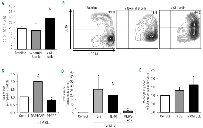
Figure 5.
In vitro CLL-induced modifications of the monocytic population. CD14+ monocytes collected from healthy donors were cultured in the presence of CLL-derived conditioned media (CM CLL) or in direct contact with CLL cells or normal B cells. (A) Histograms represent the percentage of CD16+ cells in the monocytic population collected from healthy donors (n=8) measured by flow cytometry at baseline and after 24 h-culture with normal B cells or with CLL cells. Data are reported as mean±SEM of three independent experiments (*P=0.012, Wilcoxon’s test). (B) Counter plots of a representative sample showing an increase of CD16+ cells among CD14+ monocytes after CLL contact. (C-D) mRNA levels were measured by RT-PCR on normal monocytes (n=8) collected after 24 h of treatment with CM CLL and represented as fold change compared to control medium. CLL-derived soluble factors up-regulate the expression of RAP1GAP, IL-10, IL-8 and MMP9, whereas they down-regulate PTGR2. All *P<0.05 compared to untreated controls (Wilcoxon’s test). (E) CM-CLL stimulates migration of normal monocytes. Monocytes were induced to migrate through a 5 μm-pore size PET membrane for 2 h in response to Iscove’s modified Dulbecco’s medium (IMDM) (negative control) or 10% fetal bovine serum (FBS) (positive control), and with addition of CM-CLL. Histograms represent mean±SEM of three independent experiments. Data are normalized on IMDM controls and represented as fold change. *P<0.05 compared to IMDM control (Wilcoxon’s test).

