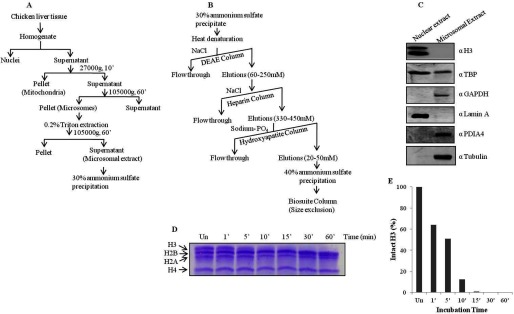
FIGURE 1.

Scheme to purify H3-clipping activity from microsomes. A, shown is a schematic of the preparation of microsomal extract from chicken liver tissue. B, shown is a scheme for the purification of H3-clipping protease from microsomal extract. C, Western blot analysis determined the purity of the microsomal extract prepared. Antibodies used for the Western blot are indicated on the right of the respective figure panels. D and E, shown is a time point assay of protease activity purified according to the above scheme and its quantification.
