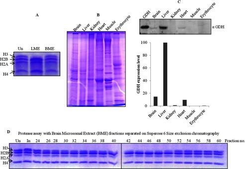
FIGURE 12.

Tissue-specific expression of GDH. Un stands for undigested core histones. A, shown is protease activity of GDH in chicken liver (LME) and brain (BME) microsomal extracts incubated for 1 h at 37 °C and analyzed on 15% SDS-PAGE. B, whole cell extracts were prepared from various tissues (indicated on top of the gel) were run on 10% SDS-PAGE. C, an immunoblot analysis shows the presence of GDH. Equal amounts of whole cell extracts were transferred and immunoblotted using anti-GDH (anti-GLUD1) antibody to determine the expression of GDH in the different tissues (upper panel). The GDH present in the tissues was then quantified (lower panel). D, brain microsomal extract was fractionated by Superose 6 size exclusion chromatography. Protease activity in fractions, noted above the gel lanes, was determined by incubation with core histones for 4 h at 37 °C.
