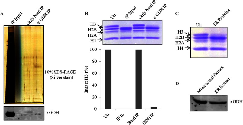
FIGURE 4.
Immunoprecipitation of GDH. A, immunoprecipitation (IP) fractions eluted from anti-GDH antibody beads or unmodified beads as control were resolved and visualized on 10% silver-stained SDS-PAGE (upper panel). Western blot analysis of the fractions for the detection of GDH was conducted using anti-GDH (anti-GLUD1) antibody (lower panel). B, shown is a H3 protease activity assay with immunoprecipitation fractions and quantification of the activity. In, input. C, shown is thr presence of protease activity in the ER. A small volume from the fractions containing ER proteins was incubated with brain core histones that were then resolved by 15% SDS-PAGE. Un, undigested core histones. D, shown is an immunoblot analysis of microsomal and ER extract using anti-GDH antibody.

