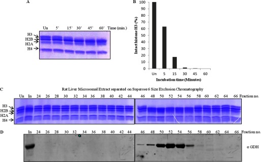
FIGURE 5.

Rat liver GDH has histone H3-clipping activity. A, shown is a time point assay with microsomal extract from rat liver. The extract prepared from the liver tissue was incubated with core histones at 37 °C for different durations (minutes of incubation are shown on the top of the gel). Un, undigested, denoting undigested core histones. The reaction mixtures were resolved on 15% SDS-polyacrylamide gel. B, shown is quantification of protease activity from rat liver. The protease activity of rat liver microsomal extract was quantified in terms of the amount of intact H3 present after the assay in A. C and D, shown is coelution of protease activity and GDH. Partial purification of protease activity of GDH from the rat liver microsomal extract was performed. The microsomal extract from rat liver tissue was subjected to size exclusion chromatography on a Superose 6 column. The chromatography fractions were assayed for the presence of the histone H3-specific tail-clipping activity (C) and GDH by immunoblotting using anti-GDH (anti-GLUD1) antibody (D). In, input.
