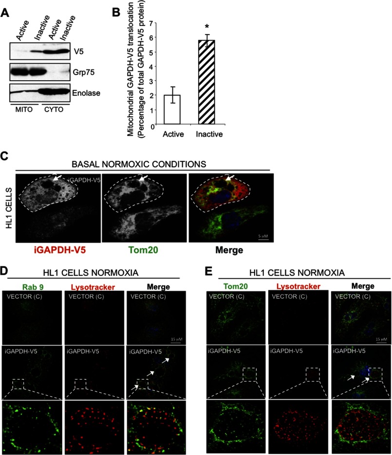
FIGURE 4.
Exogenously expressed iGAPDH associates with mitochondria and induces the formation of LL structures under normoxic condition. A, representative Western blot showing that inactive GAPDH preferentially associates with mitochondria. HL1 cells transiently expressing GAPDH-V5 (active) or iGAPDH-V5 (inactive) under normoxic conditions were fractionated for mitochondrion (Mito)- and cytosol (Cyto)-enriched fractions and analyzed by Western blotting with anti-GAPDH. Grp75 and enolase were used as markers of mitochondrial and cytosolic fractions, respectively. B, quantification of GAPDH translocation experiments described in A, using ImageJ software (n = 4, *, p < 0.05). C, HL1 cells transiently expressing iGAPDH-V5 were maintained under normoxic conditions. Cells were fixed and stained with anti-V5 (as a marker of transfected GAPDH) and anti-Tom20 antibodies (as a marker of mitochondria). Confocal images were obtained at ×100 magnification. Arrows indicate the presence of LL structures. D and E, HL1 cells transiently expressing iGAPDH-V5 or control vector (c) were labeled with LysoTracker Red, fixed, and analyzed by immunofluorescence with anti-Rab9 (D) and with anti-Tom20 (E). Images were acquired at ×100 magnification. LL structures are indicated by white arrows.

