FIGURE 1.
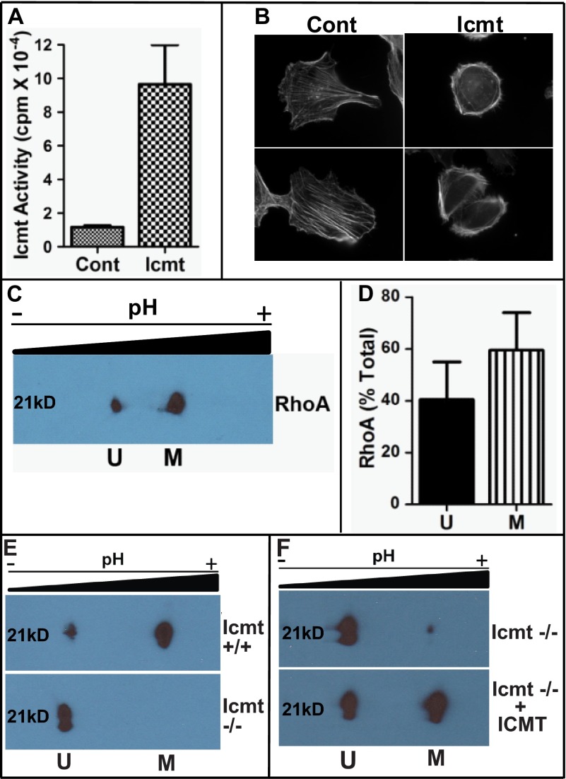
Overexpression of Icmt impacts cell morphology. A, increased Icmt enzymatic activity upon overexpression in MDA-MB-231 cells. Cells expressing GFP (Cont (control)) of GFP-Icmt (Icmt) were harvested, membranes were isolated, and Icmt activity was determined as described under “Experimental Procedures.” Data are shown as the mean ± S.E. (n = 4 for each condition). B, visualization of actin cytoskeletal structure in MDA-MB-231 cells in the presence of increased Icmt activity. Cells were plated on coverslips coated with fibronectin and allowed to adhere overnight. Representative images are displayed showing the more rounded morphology and altered actin cytoskeletal structure in GFP-Icmt-overexpressing cells (Icmt) as compared with cells only expressing GFP (Cont (control)). C, two-dimensional gel analysis of RhoA methylation status. MDA-MB-231 cells were harvested and processed, and the presence of unmethylated Rhoa (U) in the more acidic pool or methylated RhoA (M) in the more basic pool was detected by immunoblot analysis with a RhoA-specific antibody. D, quantitative representation of unmethylated and methylated RhoA levels from C, expressed as a fraction of the total amount of RhoA. Data represent the mean ± S.E. from pooled results of two independent experiments. E, validation of two-dimensional gel analysis to identify methylated and unmethylated RhoA. Cell lysates from wild-type (Icmt +/+) and null Icmt (Icmt −/−) mouse embryonic fibroblasts were harvested and processed as described under “Experimental Procedures,” and the presence of unmethylated RhoA (U) in the more acidic pool or methylated RhoA (M) in the more basic pool was detected by immunoblot analysis with a RhoA-specific antibody. F, in vitro methylation of RhoA. To confirm methylation of RhoA, recombinant Icmt was incubated with Icmt null lysate as described under “Experimental Procedures.” Icmt −/− lysate was mock-treated (Icmt −/−) or incubated with recombinant Icmt (Icmt−/− + ICMT) prior to two-dimensional gel analysis.

