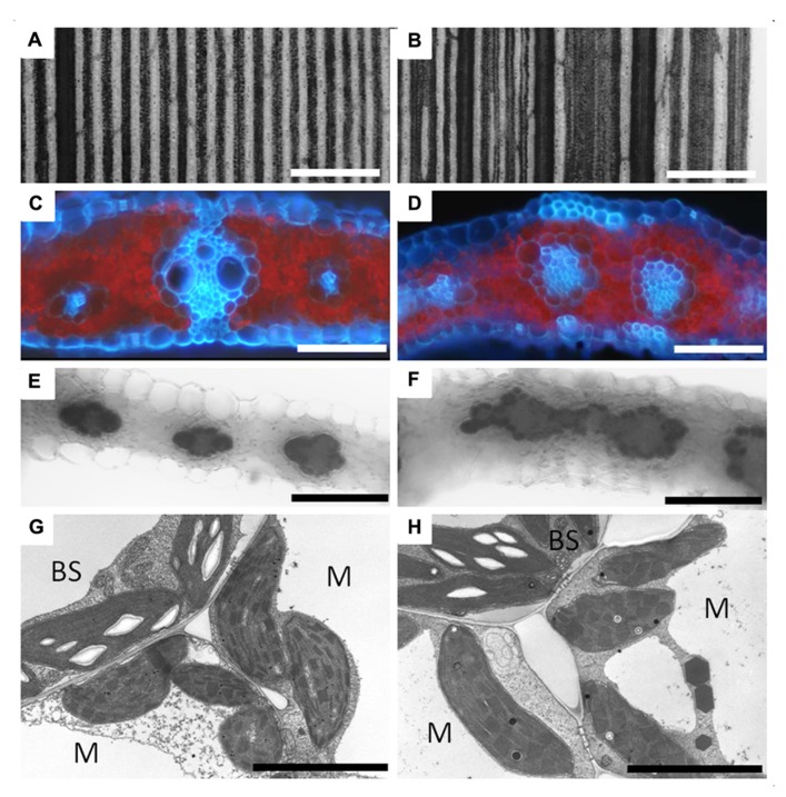
FIGURE 4.
Vascular development and bundle sheath formation in the lsn mutant of maize. Panel showing wild type (A,C,E,G) and lsn mutant (B,D,F,H) maize leaf sections. (A) Section of iodine potassium iodide (IKI) stained wild type leaf showing regular and uniform vascular patterning. (B) Section of IKI stained lsn mutant leaf showing distorted vascular pattering. (C) Cross section of wild type leaf under UV light showing canonical Kranz anatomy (red represents chlorophyll autofluorescence, blue represents autofluorescence of the cell walls). (D) Cross section of the lsn mutant under UV light, showing distorted veins with internal vascular hypertrophy and irregular internal differentiation surrounded by single layers of bundle sheath and mesophyll cells. (E,F) Cross sections of IKI stained leaves, respectively, showing normal starch accumulation in the BS cells and absence of staining in the M cells. (G,H) Transmission electron micrographs of wild type (G) and lsn mutant (H) BS and M cells showing normal C4 plastid differentiation and identity in both. BS, bundle sheath cell, M, mesophyll cell, scale bars: (A,B) 600 μm; (C–F) 50 μm; (G,H) 5 μm.

