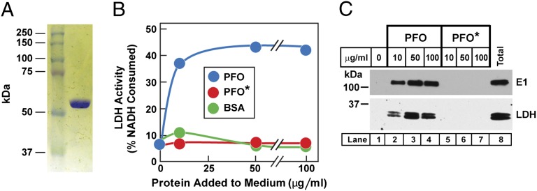
Fig. 1.
Biochemical characterization of PFO*. (A) SDS/PAGE of purified recombinant PFO*. Protein was purified as described in Materials and Methods. An aliquot (1 μg) was subjected to 8% SDS/PAGE, and proteins were visualized with Coomassie Brilliant blue R-250. (B) Lysis of SV-589 cells by PFO, but not by PFO*, as measured by release of LDH. On day 0, cells were set up in 12-well plates as described in SI Materials and Methods. On day 3, each monolayer received 0.5 mL PBS containing varying amounts of the indicated protein. After incubation for 1 h at 4 °C, the PBS was removed and assayed for LDH as described in Materials and Methods. (C) Immunoblot analysis of two cytosolic proteins released from SV-589 cells after incubation with varying amounts of purified PFO, but not PFO*. Aliquots of the cell-free PBS solution from B (lanes 1–7) and from the total cell lysate (lane 8) were immunoblotted with antibodies directed against the ubiquitin activating enzyme E1 and LDH. Before immunoblot analysis, the total cell lysate was adjusted to the same volume as each cell-free PBS solution. Blots were exposed to film at room temperature for 30–60 s.

