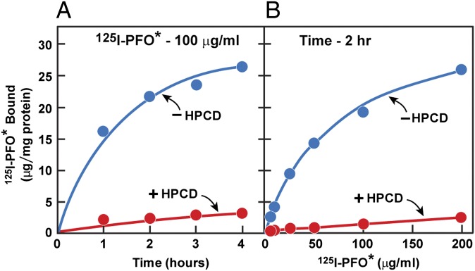
Fig. 2.
Binding of 125I-PFO* to SV-589 cells. On day 0, cells were set up in medium A at 1 × 10⁵ cells per 60-mm dish. On day 3, cells were refed with medium A. On day 4, cells were treated with fresh medium B with or without 2% (wt/vol) HPCD for 1 h at 37 °C, after which the cells were washed five times as described in Materials and Methods and then incubated at 4 °C for the indicated time with 2 mL of ice-cold buffer E containing either 100 μg/mL 125I-PFO* (6.5 × 10³ cpm/μg protein) (A) or for 2 h with the indicated concentration of 125I-PFO* (10 × 103cpm/μg) (B). The total amount of 125I- PFO* bound to the cells was determined as described in Materials and Methods. Each value represents the average of duplicate incubations. The values (mean ± SEM) for total cellular protein content did not differ significantly in cells treated with or without HPCD (0.59 ± 0.02 and 0.56 ± 0.02 mg per dish, respectively).

