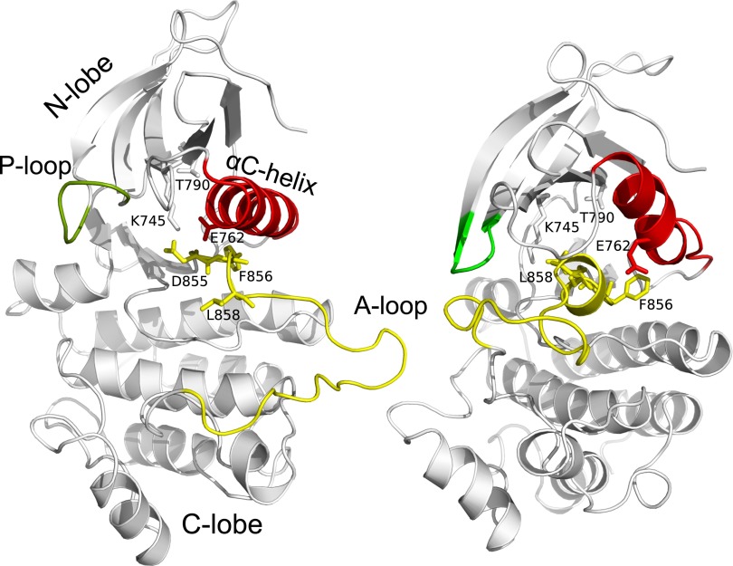
Fig. 1.
Comparison of active (Left) and Src-like inactive (Right) structures of EGFR CD. Key structural elements are colored in red (αC helix), green (P-loop) and yellow (A-loop). Also shown as sticks is the DFG motif and the residues either mutated (L858, T790) or participating in salt-bridge interactions and used to define a collective variable (K745, D855, E762).

