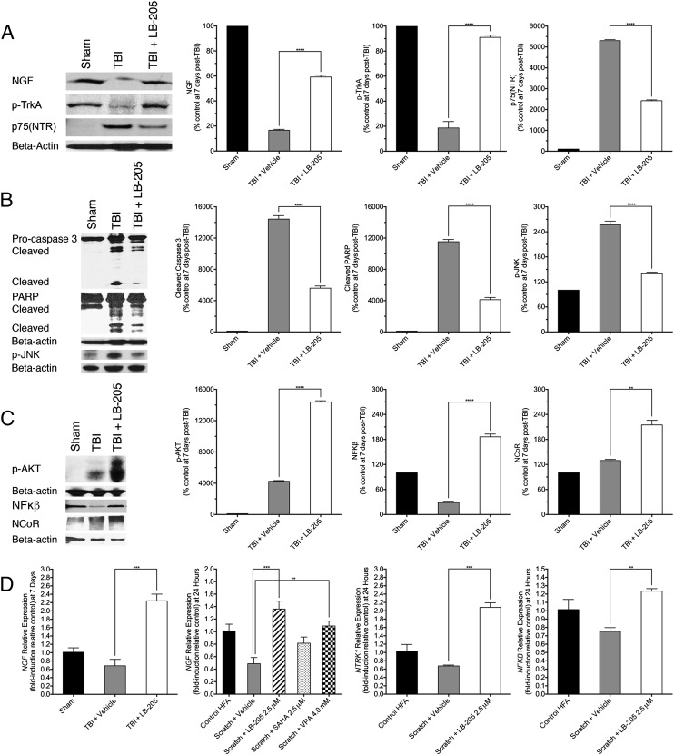
Fig. 4.
LB-205 administration preserves NGF TrkA pathway activity post-TBI in vivo and after astrocyte scratch in vitro. (A) Western blot analysis demonstrated significantly increased expression of NGF and p-TrkA and attenuated p75(NTR) expression following LB-205 treatment at 7 d post-TBI compared with TBI alone. Data are presented as mean ± SEM (n = 3; ****P ≤ 0.0001). (B) Western blot analysis indicated significantly diminished expression of p-JNK, cleaved caspase-3, and PARP following LB-205 treatment post-TBI compared with TBI alone. Data are presented as mean ± SEM (n = 3; ****P ≤ 0.0001). (C) Western blot analysis demonstrated significantly increased expression of p-AKT, NF-κB, and NCoR following LB-205 treatment post-TBI compared with TBI alone. Data are presented as mean ± SEM (n = 3; **P ≤ 0.01 and ****P ≤ 0.0001). (D) Quantitative real-time PCR, with expression of factors being conveyed as fold-induction relative to control sample expression (fold-induction = 1), demonstrated 1.56-fold increased expression of NGF following LB-205 treatment post-TBI in comparison with TBI alone in vivo, and 0.87-, 1.40-, and 0.48-fold increased expression in NGF, NTRK1, and NFKB at 24 h post-scratch in vitro in comparison with scratch alone. Data are presented as mean ± SEM (n = 3; **P ≤ 0.01 and ***P ≤ 0.001).

