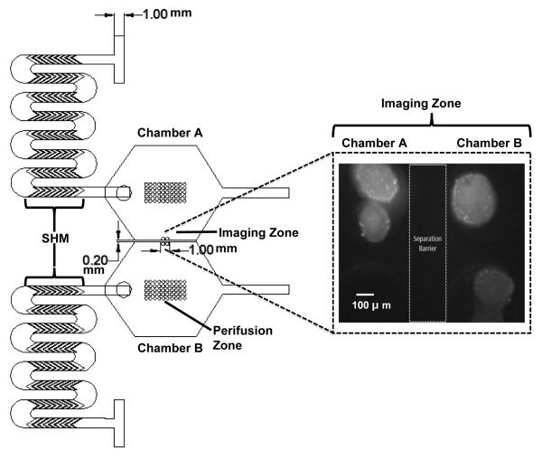
Fig. 1. A 2-D design rendering and dimensions of the dual perifusion platform.
In each perifusion network, the top-layer consists of two inlets, SHMs, a loading port, and one outlet. The middle-layer consists of a hexagonal-shape perifusion chamber. The bottom-layer has two zones of circular wells, perifusion zone and imaging zone, for islet immobilization and imaging. The image insert shows the black and white converted image of Rh123 labeled islets sitting in the imaging zones of chambers A and B, separated by a thin barrier. Scale bar = 100 μm.

