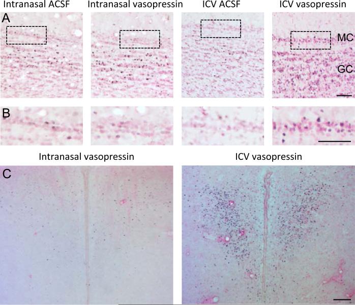
Figure 1.
Representative examples showing immunohistochemistry for the c-Fos protein in (A,B) the mitral and granule cell layer of the main olfactory bulb and (C) the paraventricular nucleus of the hypothalamus in response to intranasal or icv administration of vasopressin. MC - mitral cell layer, GC = granule cell layer. All scale bars 100 μm.

