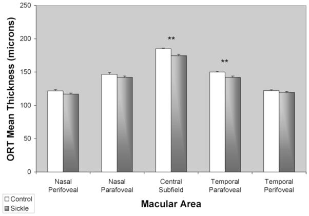
Figure 5.
Mean outer retinal thickness (ORT) in healthy control subjects (white bar) and sickle cell patients (gray bar). Thickness was averaged among the central subfield (central 1mm diameter), parafoveal (0.5 to 3 mm eccentricity) and perifoveal (3 mm to 6 mm eccentricity) regions. Error bars represent standard error of the means. (**) denotes p < 0.01.

