Figure 2.

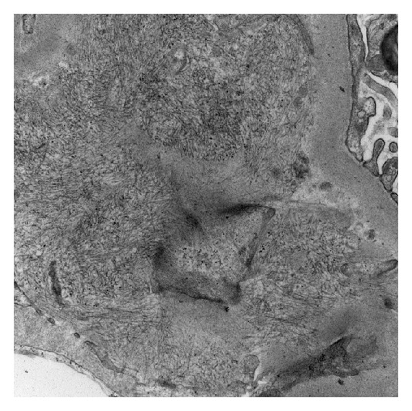
Electron micrograph showing an accumulation of randomly oriented, 18 nm in diameter fibrils in the subendothelial space between the lamina densa (upper right) and the endothelial cell (bottom left). Magnification: 20,000x.

Electron micrograph showing an accumulation of randomly oriented, 18 nm in diameter fibrils in the subendothelial space between the lamina densa (upper right) and the endothelial cell (bottom left). Magnification: 20,000x.