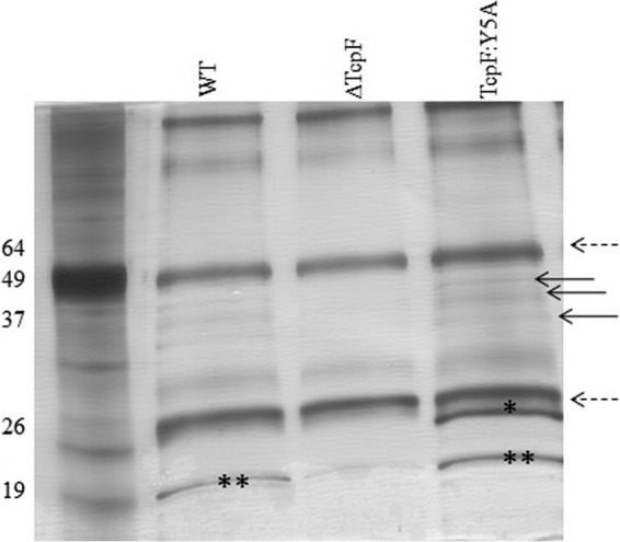
Fig 5.

Immunoprecipitation of cross-linked whole-cell lysates with anti-TcpF antibody. After immunoprecipitation, antibody-bound proteins were loaded onto an SDS-PAGE gel and subjected to electrophoresis. Proteins in the gel were silver stained. The dashed arrows correspond to heavy- and light-chain immunoglobulin. The bands with the asterisks were removed and subjected to trypsin digestion and mass spectrometry analysis. “*” corresponds to VCO395_A0815; “**” corresponds to TcpA. The arrows point to TcpF and its degradation products. WT, wild type.
