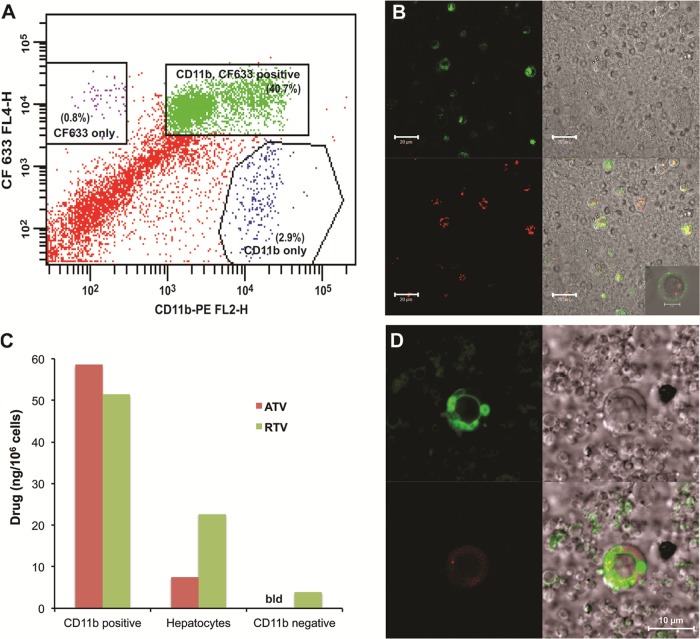
Fig 7.
In vivo colocalization of nanoART in CD11b-positive cells of the liver and storage in nonlysosomal compartments. Male BALB/cJ mice were treated SC with 250 mg/kg ATV/RTV (1:1 drug ratio) coated with CF633-modified P188. Liver cells were isolated 24 h later by in situ collagenase digestion. (A) FACS analysis of liver nonparenchymal cells from CF633-P188-ATV/RTV-treated mice incubated with CD11b antibody and collected using MACS cell separation columns. (B) Confocal microscopy of nanoART-loaded nonparenchymal cells following CD11b-positive cell purification showing localization of nanoART (red) in CD11b-positive cells (green) (bar = 20 μm; inset bar = 5 μm). (C) ATV and RTV levels in various liver cell types following cell separation using differential centrifugation and CD11b-positive MACS cell separation (data from a representative experiment are shown). Drug levels were quantitated by LC-MS/MS (bld = below detection limit). (D) Confocal microscopy of nanoART-loaded (red) nonparenchymal cells incubated with Lysotracker Green showing localization of nanoART outside lysosomal (green) compartments (bar = 10 μm).

