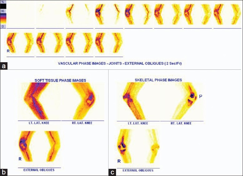
Figure 2.

Display of a regional three phase bone scan (TBS) that was performed with 740 MBq of 99mTc MDP given as IV. (a) Immediate dynamic, (b) soft tissue phase and (c) 3 hours later skeletal phase images of both knee joints were acquired using high resolution collimators on a dual head variable angle Gamma camera. Note the technical modification in limb positioning for simultaneous assessment of patellae (Annotation P denotes patella), i.e. limbs are adducted, externally rotated with 30° flexion of the knees to separate the patella from the femoral condyle)
