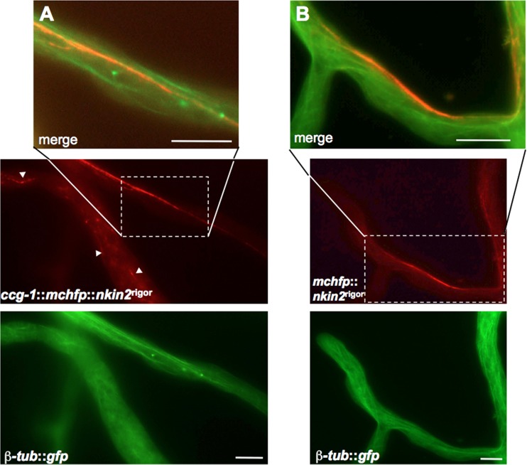
Fig 9.
Colocalization of mChFP-NKIN2rigor and β-tubulin-GFP. (A) Whereas in distal parts of primary hyphae, often only short pieces of the MT network are labeled (arrowheads) by mChFP NKIN2rigor, in lateral branches, one MT bundle appears to be preferred (magnification). (B) In apical branches of leading hyphae, GFP-NKIN2rigor decorates an MT subpopulation close to the edge of the hyphae, whereas the other MTs are not affected (magnification). Scale bars, 10 μm.

