
Fig 1.
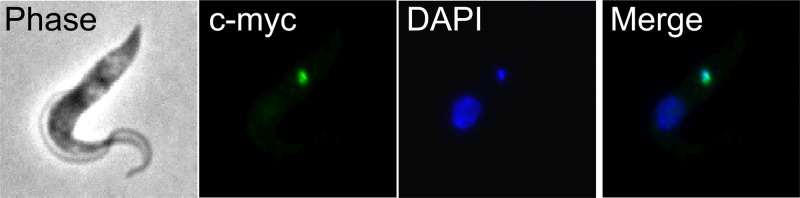
Tb927.2.6100 associates with the kDNA. The tagged protein was visualized by fluorescence microscopy using polyclonal anti-c myc antiserum coupled with fluorescein isothiocyanate (FITC)-conjugated secondary antibody. Panels show phase-contrast light microscopy of PF T. brucei cells, anti-c-myc antibody coupled with FITC-conjugated secondary antibody showing localization of the target protein, DAPI staining of nucleus and kDNA, and merge.