
Fig 4.
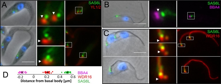
TbSAS6L-YFP localizes to a prominent focus near the base of the flagellum in T. brucei. (A) TbSAS6L-YFP (green) is distal to the basal body, which is marked by tyrosinated α-tubulin (YL1/2, red, arrowheads). A lower-intensity SAS6L-YFP signal is also associated with the developing probasal body. SAS6L-YFP is also distal to the basal-body markers BBA4 (B) and WDR16 (C) (arrowheads). (D) Distance measurements in relation to YL1/2 indicate that SAS6L-YFP localizes to a region near the axoneme basal plate (20 measurements for each marker). Scale bars, 5 μm.