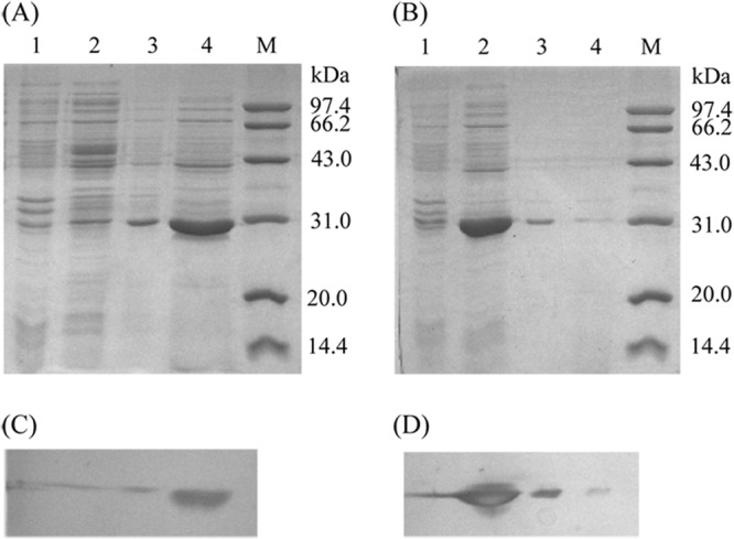
Fig 2.

SDS-PAGE (A and B) and Western blot (C and D) analysis of the enzyme (cutinaseNS [A and C] and cutinaseNSS130A [B and D]) distribution. The primary antibody was anti-six-histidine mouse IgG, and the second was HRP-labeled goat anti-mouse IgG(H+L). Lanes: M, molecular mass standard protein; 1, intracellular insoluble fraction; 2, intracellular soluble fraction; 3, periplasmic fraction; 4, culture supernatant.
