Fig 1.

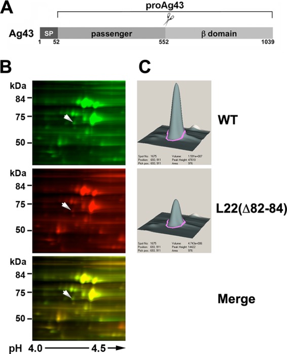
Level of Ag43 is reduced in an L22(Δ82-84) mutant strain. (A) Illustration of Ag43 showing the signal peptide (SP; residues 1 to 52), the passenger domain (residues 53 to 552), and the β domain (residues 553 to 1039). The pro form of the protein contains covalently linked passenger and β domains and is observed prior to the secretion and proteolytic processing of the passenger domain. (B) Soluble proteins from MG1655 and MNY15 [MG1655 rplV(Δ82-84)] were labeled with the fluorescent dyes Cy3 and Cy5, respectively, and analyzed by 2D-DIGE. Images of a section of each gel run in a representative experiment and an overlay image are shown. The spot that corresponds to Ag43 is denoted by an arrow. (C) Quantitative DeCyder analysis of the spot indicated in panel B reveals a 2.7-fold reduction of Ag43.
