
Fig 2.
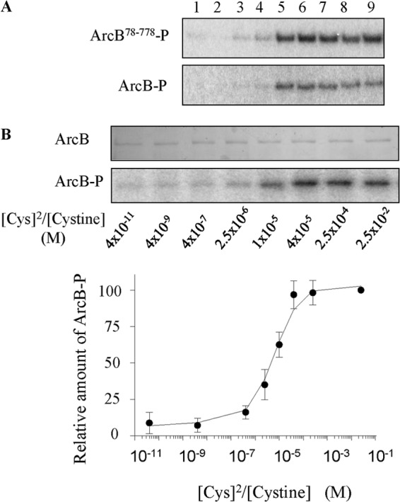
Determination of the redox potential of ArcB. (A) Purified ArcB78-778 (top) or full-length ArcB-enriched inverted membrane vesicles (bottom) were incubated with [γ−32P]ATP in the presence of 1 mM: lane 1, 1,4-benzoquinone (E′° = +274 mV); lane 2, 1,2-naphtoquinone (E′° = +134 mV); lane 3, 1,4-naphtoquinone (E′° = +69 mV); lane 4, juglone (E′° = +30 mV); lane 5, menadione (E′° = ±0 mV); lane 6, plumbagin (E′° = −29 mV); lane 7, lawsone (E′° = −137 mV); lane 8, anthraquinone-2-sulfonate (E′° = −225mV); and lane 9, nothing. After 2.5 min, the reactions were terminated by addition of an equal volume of 4× SDS sample buffer and the mixtures were immediately subjected to SDS-PAGE. Representative autoradiograms of the dried gels are presented. (B) A concentration of ∼1 μM full-length ArcB embedded in inverted membrane vesicles was incubated with buffers containing various cysteine and cystine concentrations for 3 h at 25°C. Subsequently, the protein was incubated with [γ−32P]ATP for 2.5 min, the reactions were terminated by addition of an equal volume of 4× SDS sample buffer, and the mixtures were immediately subjected to SDS-PAGE. Gel stained with Coomassie blue (top), an autoradiogram of phosphorylated proteins on the dried gel (middle), and a plot of ArcB net phosphorylation versus [Cys]2/[Cystine] (bottom) are shown. Data represent the averages from seven independent experiments, and the standard deviation values are indicated. The plot was interpreted by the equations given in Materials and Methods, and the Keq and E′° values for ArcB were determined to be 4.2 × 10⁻⁶ M and −41 mV, respectively.