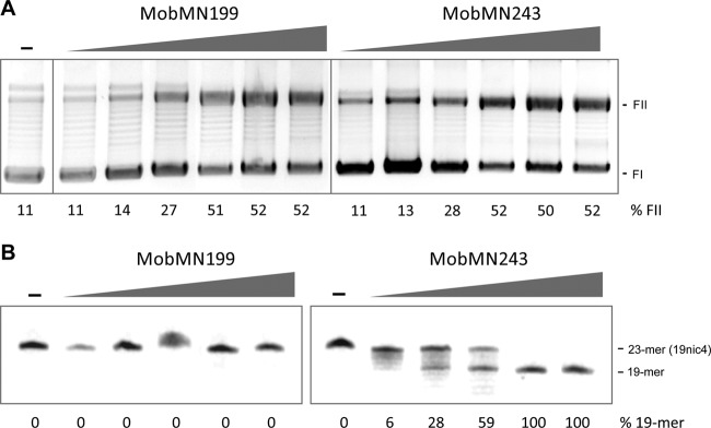
Fig 4.

Nicking activity of MobMN199 and MobMN243. (A) Relaxation assays of pMV158 by MobMN199 and MobMN243. Supercoiled pMV158 DNA samples (8 nM) were incubated with (8, 16, 32, 64, 128, or 256 nM) or without (−) MobMN199 or MobMN243 and supplemented with 8 mM MnCl2 at 30°C for 20 min. Generation of relaxed forms (FII) from scDNA forms (FI) was analyzed by electrophoresis on 1% agarose gels stained with ethidium bromide (1 μg/ml). The weak band above relaxed forms (FII) has been observed before (20) and might correspond to relaxed DNA dimers. (B) Nicking activity of MobMN199 and MobMN243 on single-strand DNA harboring regions of the pMV158 oriT (19nic4). The nicking reaction yields 19- and 4-mer (not shown) oligonucleotide products. 19nic4 (60 nM) was incubated with a range of protein concentrations (60, 120, 240, 480, and 960 nM) at 30°C for 20 min in the presence of 8 mM MnCl2. The reaction was stopped with 0.1% SDS and proteinase K (1 mg/ml) for 30 min at 37°C, and samples were electrophoretically analyzed in a denaturing 20% PAA–7 M urea gel and visualized by a phosphorimager. Band quantification was done using the QuantityOne software (Bio-Rad) for both panels.
