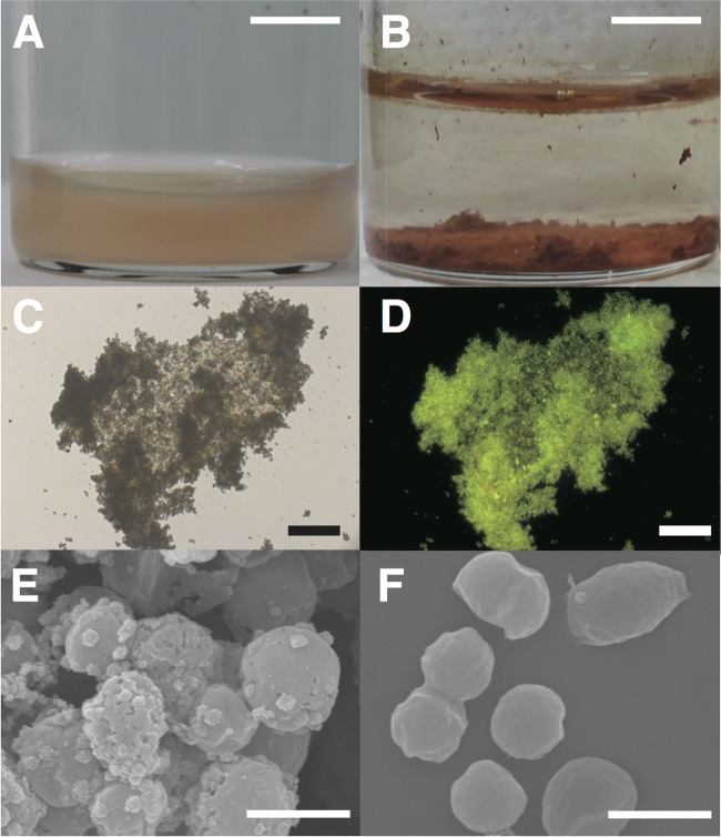
Fig 3.
Microscopy of rusty precipitates developed during nitrate-dependent Fe2+ oxidation by “Ca. Brocadia sinica.” Cell suspensions of “Ca. Brocadia sinica” were anaerobically incubated with NO3− and Fe2+ at 37°C (A). Rusty precipitates exhibiting a reddish color appeared at the bottoms of the vials after 1 day of incubation (B). Precipitates were stained with SYBR gold and subsequently examined by epifluorescence microscopy. Precipitates gave a signal for SYBR gold. Panels C and D are bright-field and fluorescence microscopy images, respectively, of SYBR gold. The precipitates were further examined by SEM (E). Cells were covered with small amorphous particles and were attached via the particles. As a control, “Ca. Brocadia sinica” cells were examined prior to incubation (F). Amorphous particles were not observed in image F. Scale bars are 6 cm (A and B), 100 μm (C and D), and 1 μm (E and F).

