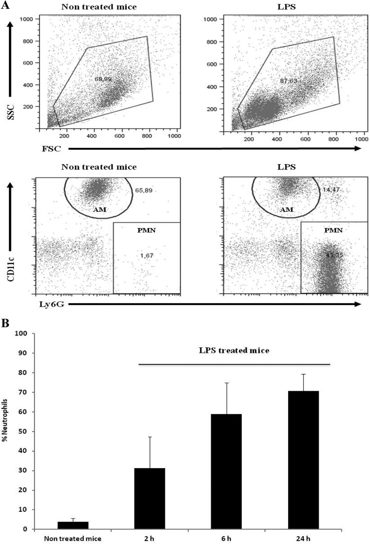
Fig 2.
Leukocyte recruitment into the airways after LPS treatment. The cellular populations in the airways were sampled by BAL and analyzed by flow cytometry at the indicated time points. (A) BAL fluid dot plots representative of PBS-treated mice (nontreated) and LPS-treated mice. The diagram corresponds to BAL fluid obtained 24 h after treatment. Alveolar macrophages (AM) were defined as CD11c+ CD11b+ Ly6G− GR1− cells, and neutrophils (PMN) were defined as CD11c− CD11b+ Ly6G+ GR1+ cells. (B) Quantitative analysis at different times after treatment of BAL fluid PMN obtained from PBS-treated animals (nontreated) and LPS-treated mice.

