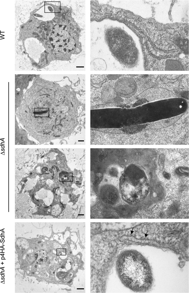
Fig 3.
LCVs of L. pneumophila ΔsdhA in hemocytes, showing signs of instability. G. mellonella larvae were infected with WT, ΔsdhA, or ΔsdhA plus p4HA-SdhA L. pneumophila 130b for 5 h. Hemocytes were extracted and processed for transmission electron microscopy. Hemocytes infected with WT bacteria contained LCVs of similar morphology to those previously described. Recruitment of the ER to the LCV is indicated (black arrows). The LCVs of the ΔsdhA mutant contained electron-dense material, which may be cytoplasmic or bacterial in origin, indicating loss of stability of the vacuole. The LCVs of the complemented strain were of comparable morphology to those of WT bacteria. Images are representative of two independent experiments. Bar, 1 μm.

