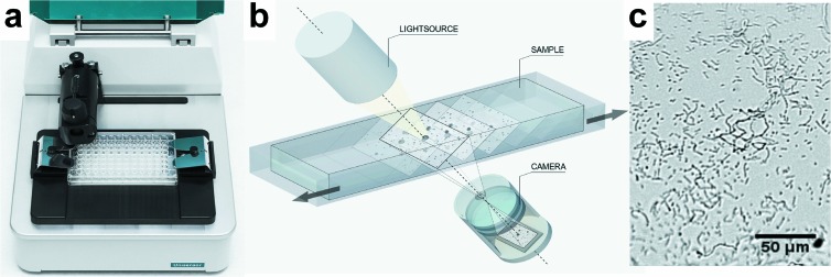
Fig 1.
The oCelloScope detection system. (a) Picture of the detection system with a standard 96-well plate inserted. (b) Simplified engineering drawing of the detection principle. A volume of 50 μl of a growing bacterial culture is scanned, resulting in a stack of images. Each image can be transformed to a two-dimensional (2D) picture. (c) 2D picture of S. alactolyticus from the scanning process. The magnitude is comparable to an ×200 magnification in a standard light microscope.

