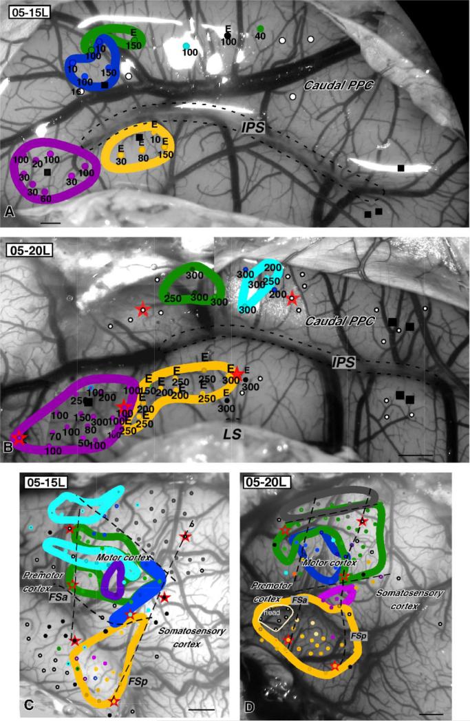
Figure 5.
The organization of PPC in cases 05-15L (A) and 05-20L (B). Sites and zones where complex forelimb and face movements were evoked from motor cortex are marked in both cases on the dorsolateral surface of left hemisphere (C,D). In both cases, regions of primary motor cortex and premotor cortex were stimulated with long stimulus trains, evoking complex face, forelimb, and hindlimb movements similar to those evoked from PPC. Color code (see previous legends) is the same as for PPC. bi, Bilateral; E, ear; ext, extension; fl, forelimb; FSa, anterior frontal sulcus; FSp, posterior frontal sulcus; LL, lower lip; M, mouth; T, tongue; Tr, trunk. Conventions as in Figures 2 and 3. Scale bars = 1 mm.

