
Figure 2.
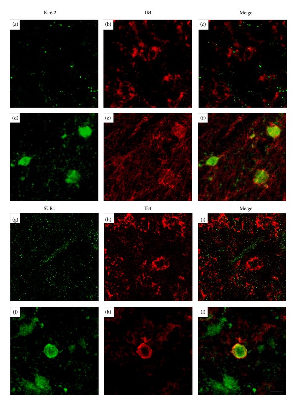
Expression of KATP channel components SUR1 and Kir6.2 in activated IB4-positive cells into the core of the hippocampal lesion (Bregma −3.3). ((a)–(f)) Confocal photomicrographs of hippocampal sections immunostained with IB4 and anti-Kir6.2 antibodies in control ((a)–(c)) and NMDA-lesioned rats ((d)–(f)). ((g)–(l)) Confocal photomicrographs of hippocampal sections immunostained with IB4 anti-SUR1 antibodies in control ((g)–(i)) and NMDA-lesioned rats ((j)–(l)). Note that reactive amoeboid microglia stained with IB4 show specific immunostaining with anti-Kir6.2 and anti-SUR1 antibodies in the cell membrane but also in the cytoplasm. For lesion details, see legend of Figure 1. Scale bar 10 μm.