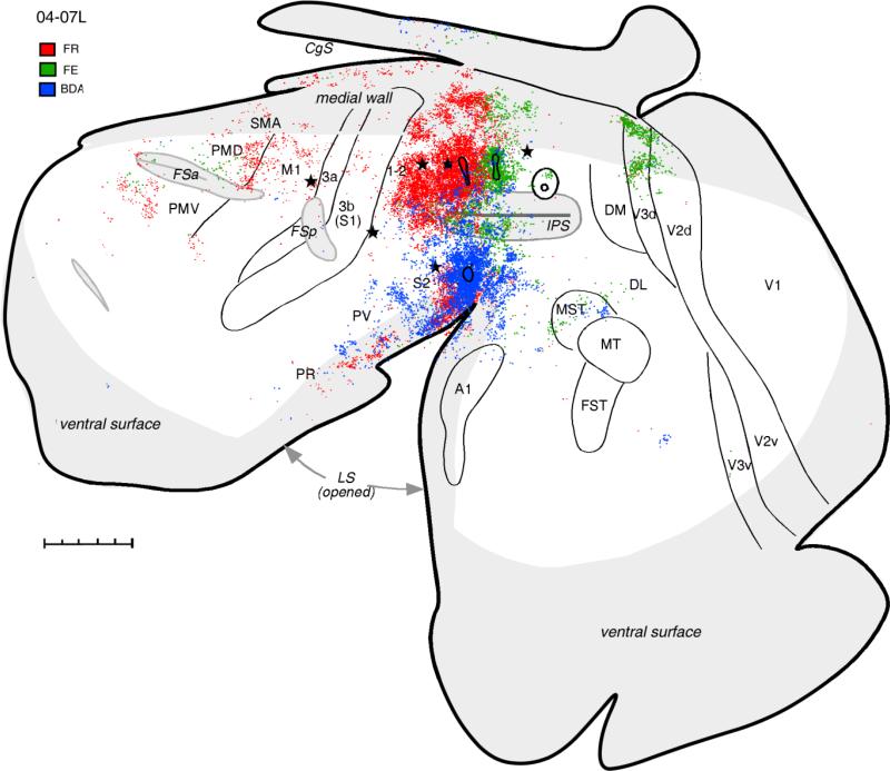
Figure 5.
Distributions of labeled neurons after injections of tracers FR (red dots), FE (green dots), and BDA (blue dots) in different locations in anterior PPC in case 04-07L. The pattern of connections after the FR injection that partially covered hand-to-mouth and defensive forelimb zones is similar to other cases with hand-to-mouth zone injections, although less label was obvious in M1 in this case. Note also the presence of label in the extrastriate visual area V3d. FE injection in the forelimb defensive zone labeled the most cells in adjacent parts of PPC and in extrastriate visual areas. Fewer cells were labeled in the premotor and cingulate cortex, and only few in the somatosensory areas. Injection in the face defensive zone labeled the most neurons in the somatosensory areas of the lateral sulcus and in the auditory belt and visual area MST. Injection in posterior PPC is shown for reference. For the detailed microstimulation map of this case see Figure 2B in our companion paper. Conventions as in Figure 2. Scale bar = 5 mm.

