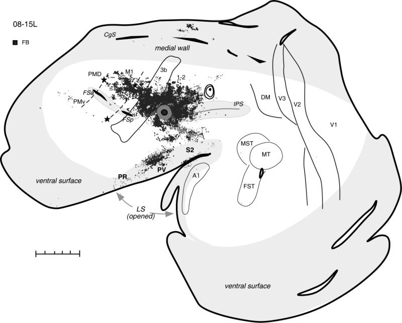
Figure 8.
Distributions of labeled neurons after injection of FB in the most rostral part of PPC responsive to ICMS in case 08-15L. In this case, most labeled cells were found in the somatosensory areas of anterior parietal cortex and in the lateral sulcus, as well as in the motor cortex. Premotor and cingulate cortex had fewer labeled cells, and visual areas were free of label. For the detailed microstimulation map of this case see Figure 8A. Conventions as in Figure 2. Scale bar = 5 mm.

