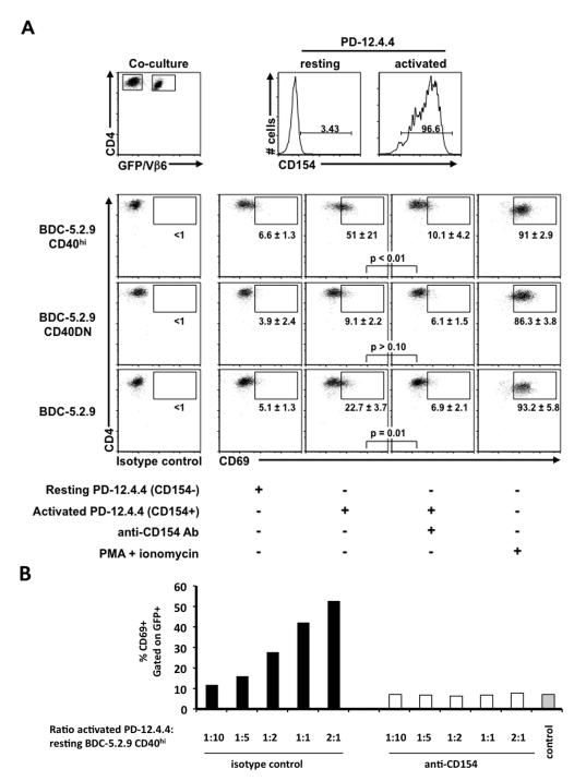
Figure 2.
CD154-positive T cells can activate CD40-positive T cells. The insulin-reactive T cell clone PD-12.4.4 (Vβ6-negative) was activated with immobilized anti-CD3 (2 μg/ml). After 18 hr, the PD-12.4.4 T clone was extensively washed in CM and co-cultured 1:1 (or at the ratios indicated) with 1 × 10⁵ resting T cell clones: BDC-5.2.9 (Vβ6+), BDC-5.2.9 CD40hi (GFP+) or BDC-5.2.9 CD40DN (GFP+), in the presence or absence of anti-CD154 Ab (10 μg/ml) or isotype control Ham IgG. Four hours after co-culture, cells were harvested and stained with anti-Vβ6 FITC, anti-CD4 APC and anti-CD69 PE or anti-CD154 PE or appropriate isotype control. (A) Expression of GFP, Vβ6, CD154, CD69 and CD4 was monitored by flow cytometry and gates were set on live CD4 cells. CD69 expression was monitored on GFP+ cells and was comparable on all the clones when stimulated with PMA/ionomycin. (B) Gates were set on live CD4+ GFP+ cells and the percentage of CD69+ cells is represented for each condition. Black bars: co-cultures in the presence of isotype control; white bars: co-cultures in the presence of anti-CD154 blocking antibody; gray bar: co-culture of BDC-5.2.9 CD40hi with resting PD-12.4.4 (CD154-negative). Data are representative of at least three independent experiments.

