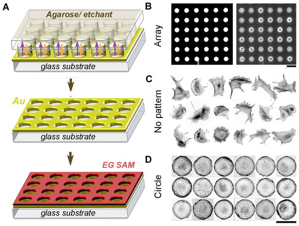
Figure 1. Control of cell geometry on transparent microislands.
(A) Scheme of the reaction-diffusion microetching (see text for description). (B) An array of microetched circular islands. Left, bright field image (white = etched islands; dark = unetched gold); right, phase-contrast image showing that cells assume the shapes of the etched islands. (C) B16F1 mouse melanoma cells on unpatterned substrate display heterogeneous cell shapes and cytoskeletal organization (cells stained with fluorescent phalloidin to visualize actin filaments, image contrast-inverted). (D) B16F1 cells on micropatterned circular islands have identical shapes and display approximately uniform organization of actin cytoskeleton; 18 different cells are shown. Scale bars are, 80 μm for B and 40 μm for C, D.

