
Figure 4. Cell spreading on circular micropatterns at early times ( t = 0–2h).
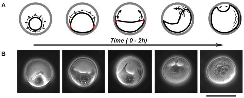
(A) Schemes depicting cell spreading on circular microislands (see text for detailed discussion). Thicker lines mark cell body; thinner lines outline protrusive lamellipodia; arrows indicate spreading/lamellipodia protrusion directions; wavy lines indicate ruffles; red marks indicate high-curvature points. (B) Microscopy images (phase-contrast) depicting the stages of cell spreading outlined in the schemes in A. Dark = unetched gold; grey = etched islands with cells. Scale bar is 40 μm.