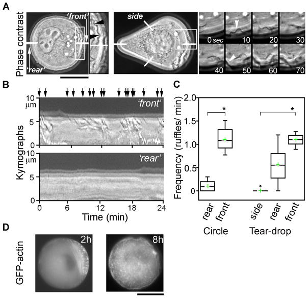
Figure 6. Time-lapse of membrane ruffling dynamics in live B16 cells on circular and teardrop shape islands.
(A) At 2h, circular (left) and tear-drop (right) shape cells display polarized/asymmetric ruffling activity--ruffling occurs predominantly at one end of the cell (here, termed “front”); ruffling activity is absent at the other end of the cell (“rear” for circles or “sides” for tear-drops, see also kymograph analysis in B). For circular cell, inset is an enlarged “front” region (indicated by white box) showing ruffles (black arrowheads); ruffles appear as dark wavy lines (low grey values). For tear-drop, expanded inset shows time-series of phase-contrast images (white arrowheads indicate appearance of two ruffles). See also Supplementary Movies 2–3 Online. Scale bar = 20 μm. (B, C) Ruffling activity quantified using kymograph analysis (see Materials and Methods). Kymographs (B) or time-space plots of intensity values in linear regions (shown as white lines in A) across the planes of the time-lapse stack. Images show typical kymograph plots for “front” and “rear” regions for circular cells (“sides” of tear-drops look similar to “rear” of circles). Ruffles are identified by dark appearance in phase-contrast (low grey values) and by their centripetal movement from the cell’s edge toward cell interior (indicated by slanted orientation of dark lines in the kymographs). Note numerous ruffles in “front” kymograph (black arrows point at individual ruffles) and their absence in “rear” kymograph. (C) Ruffles were enumerated from kymograph images of cells imaged 2–4 hrs after plating. “Front” regions display statistically significantly increased frequency of ruffles. Top and bottom of the box show 75th and 25th quartiles, respectively; whiskers indicate minimum and maximum values; circles show outliers, middle line is the median and cross is the mean. (n=11 cells; 263 minutes total observation for circles and n=7 cells, 154 min for tear-drops; *p <0.0001; Student’s t-test.) (D) The areas of polarized ruffling activity are coincident with polarized asymmetric accumulation of GFP-actin in transverse bundles and spots (2h). At 8h GFP-actin is symmetrically distributed. See also Supplementary Movie 1 Online. Scale bar = 20 μm.

