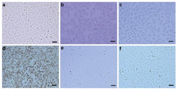
Figure 2.

Bright-field micrographs of HeLa cells (a) without treatment, treated with (b) GNR-silica-FA, (c) silica-MNP-FA, (d) GNR-silica-MNP-FA, (e) GNR-silica-MNP, and (f) GNR-silica-MNP-FA together with 1 mM free folic acid. The dark appearance of cells in (b–d) is due to nanoparticles inside cells promoted by FA receptor targeting. Quantitative assessment of non-specific trapping of cells in panel (f) using PA imaging is shown in Supporting Information Figure S3.
