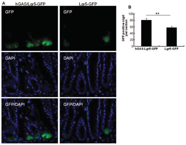
Figure 6.
Lgr5-GFP localization is increased in hGAS compared with WT mice. (A) The localization of Lgr5-GFP positive cells in hGAS/Lgr5-GFP and Lgr5-GFP mice colon. (B) The average of Lgr5-GFP positive cells in crypts in each of group were measured under a immunofluorescence microscope. We had total of three mice in each group and three sections were examined per colon. All values represent the mean ± SD. **p < .01.

