Fig. 2.
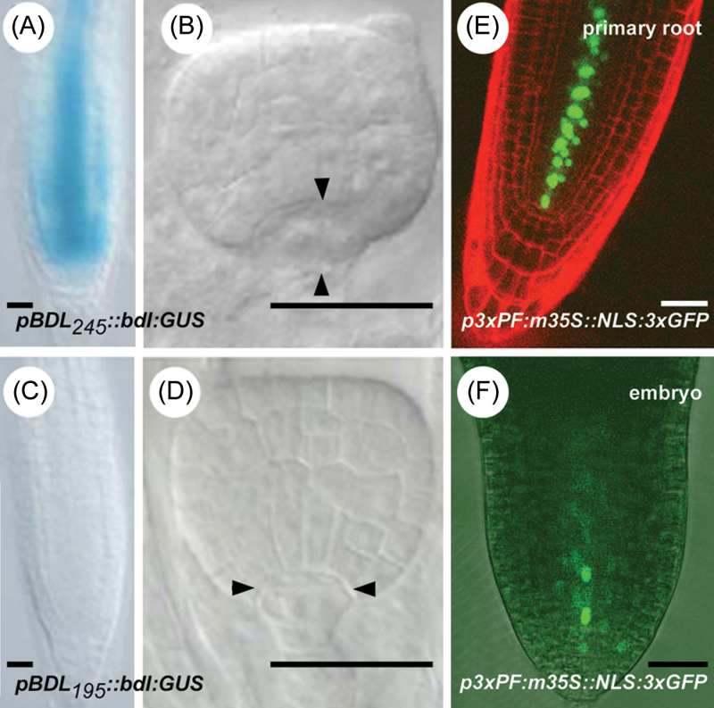
pBDL deletion study. (A) GUS-stained pBDL 245 ::bdl:GUS seedling root tip. (B) Hypophysis division defect (vertical instead of horizontal division) in pBDL 245 ::bdl:GUS embryos. (C) GUS-stained pBDL 195 ::bdl:GUS seedling root tip. (D) Normal hypophysis division in pBDL 195 ::bdl:GUS embryos. (E, F) p3×PF:m35S::NLS:3×GFP expression in a seedling root counterstained with propidium iodide (E) or in a torpedo-stage embryo (basal part shown) (F). Bars, 25 μm.

