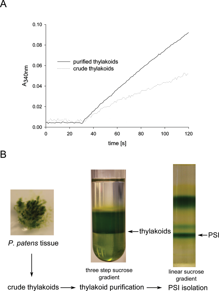
Fig. 1.
Isolation of thylakoids from Physcomitrella. (A) Absorption trace (at 340nm) of in vitro NADP+ photoreduction assay using crude thylakoids (dotted trace) or purified thylakoids (solid trace) of Physcomitrella. (B) Physcomitrella tissue was disrupted and a crude thylakoid extract prepared, which was subjected to a further clean-up step via a discontinuous sucrose gradient (1.8, 1.3, and 0.5M sucrose layered on top of each other). After centrifugation, the thylakoids were collected at the 1.8/1.3M sucrose interphase. The thylakoids were used for biochemical assays or were solubilized using β-d-dodecyl-maltoside and subjected to a centrifugation on a linear sucrose gradient to obtain intact the PSI–LHCI complex.

