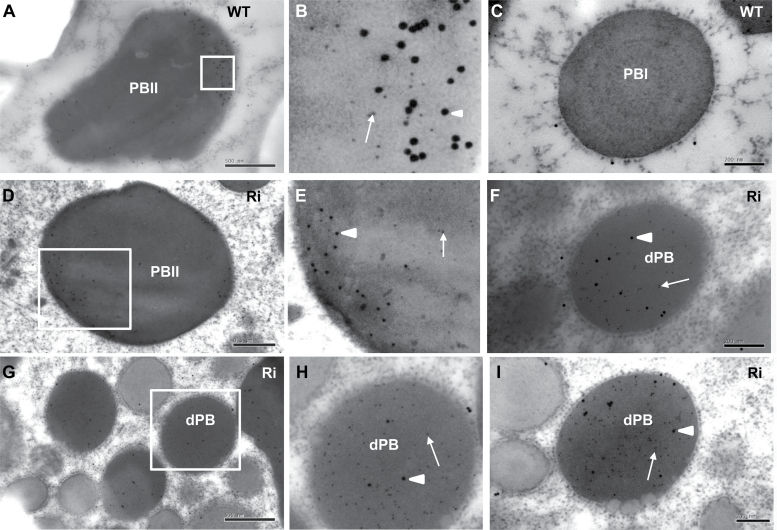
Fig. 8.
Immunocytochemistry of glutelin and α-globulin in protein bodies. (A–C) Wild type; (D–I) OsSar1abc RNAi transformants. (B, E, H) Enlarged images of areas inside the boxes in (A, D, G), respectively. Glutelin and α-globulin antibodies are labelled with 5nm (white arrow) and 15nm (white arrowhead) immunogold particles, respectively. Scale bars=500nm in (A, D, G) and 200nm in (C, F, I).

